“Tale of two supers”: super-resolution microscopy meets supervised learning
Akin to human skeleton there are molecules that polymerize to form a cellular skeleton helping the cell maintain its shape and function. These structures form the cytoarchitecture of the cells and are referred as cytoskeleton. Actin is one of the prominent molecules that form the cytoskeleton at the synapses. Synapses are micron-scale structures where the communication between neurons occurs and their structure and function are intimately associated with cognitive processes like learning and memory. The polymerized form of actin – “Filamentous actin (F-actin)” is the key organizational network forming variety of shapes and giving the three-dimensional dynamic structure for neuronal synapses. Thus, the organization of F-actin nanostructures is known to be one of the key factors in deciding the structure and function of these synapses in health. A deviation from normal function or organization of F-actin is implicated in cognitive disabilities and neurodegenerative diseases like Alzheimer’s disease. Multicolour super-resolution microscopy enables us to have better insights into the nanoscale architecture of the synapse in health and disease. Here, we have combined novel methods for analysis of nanoscale images of F-actin network using segmentation with pattern recognition based on supervised learning. A key feature of this automated paradigm is its ability to extend the shape features to include multiple classes of synaptic morphologies obtained by super-resolution images along with the nanoscale skeleton of the actin network. The automated approach was used to differentiate the heterogeneity in the nanoscale organization of F-actin in primary neuronal cultures from wild type and a transgenic mouse model of Alzheimer’s Disease. This paradigm was able to show that branched nanoscale organization of F-actin networks are altered within synaptic sub-compartments much before the onset of AD.
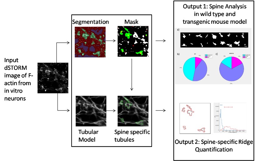
Schematic representation of the workflow for generating an objective classification of F-actin organization in dendritic spines
This project was supported by DBT, IISC and Tata Program grant towards understanding the early detection and treatment of AD
Reference : Characterization of Nanoscale Organization of F-Actin in Morphologically Distinct Dendritic Spines In Vitro Using Supervised Learning, Siddharth Nanguneri, R. T. Pramod, Nadia Efimova, Debajyoti Das, Mini Jose, Tatyana Svitkina and Deepak Nair, eNeuro 16 July 2019, 6 (4) ENEURO.0425-18.2019
DOI: https://doi.org/10.1523/ENEURO.0425-18.2019
Website URL : http://www.cns.iisc.ac.in/deepak/Homepage.html