Nanofiber platform to study muscle disorders
– Debraj Manna
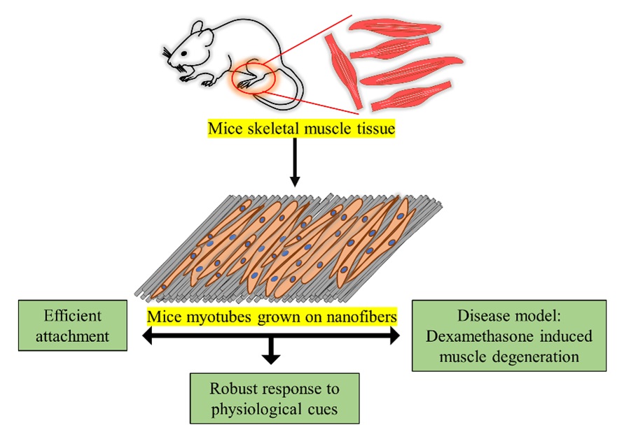
Skeletal muscles are formed by the fusion of parent muscle cells called myoblasts. These muscles are usually tethered to the bones by tendons. Because we use them to move different body parts, skeletal muscle disorders accompany loss in muscle fibers that often causes difficulty in movement, pain, and stiffness upon stress and aging.
Despite extensive research in the lab, current treatments for such disorders are often ineffective. This gap, in taking knowledge from the bench to the bedside, is mainly because of the lack of suitable lab-made muscle models that can recapitulate behaviour akin to that of skeletal muscles inside the body. Researchers at the Centre for BioSystems Science and Engineering, Department of Materials Engineering and Department of Microbiology and Cell Biology have recently addressed this issue. They used nanofibers of polycaprolactone (PCL), a biodegradable polyester, to create a mesh-like structure on which they cultured myoblasts in the lab and allowed the cells to grow into muscle fibers.
Experiments showed that the lab-grown muscle fibers retain their alignment when grown on such polymeric substrates and could reproduce critical properties of skeletal muscles, such as stress-induced muscle degeneration. The nanofiber mesh offers a robust platform to not only study muscle disorders, but also to test the effectiveness of drugs to treat them.
REFERENCE:
Aditi Jain, Manisha Behera, Venkatraman Ravi, Sneha Mishra, Nagalingam R. Sundaresan, Kaushik Chatterjee, Recapitulating pathophysiology of skeletal muscle diseases in vitro using primary mouse myoblasts on a nanofibrous platform, Nanomedicine: Nanotechnology, Biology and Medicine (2021).
https://www.sciencedirect.com/science/article/pii/S1549963420301957
LAB WEBSITES:
http://sundaresanlabiisc.weebly.com/
https://sites.google.com/site/iiscbiomaterials/